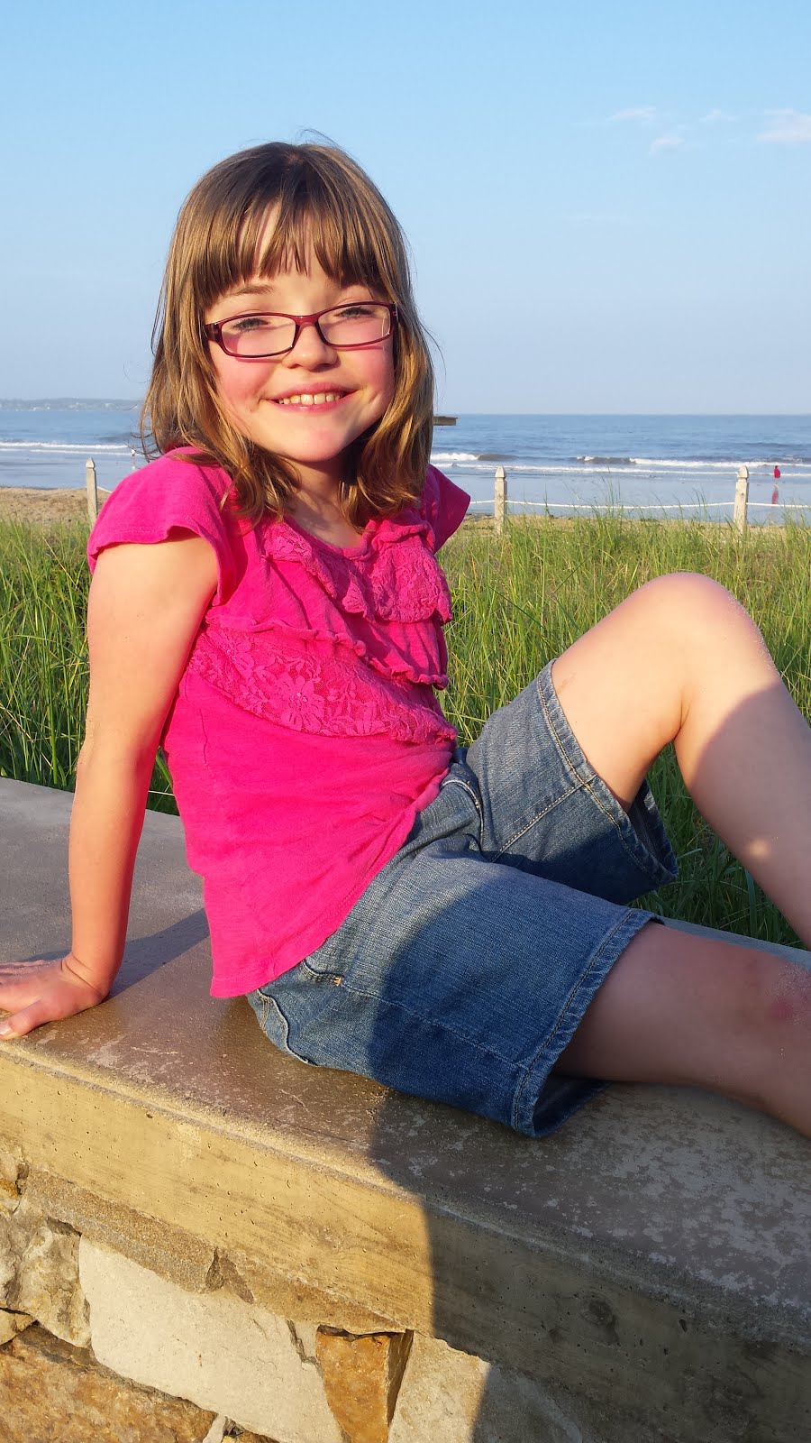
Today was busy here. Grandma kindly came to pick Jason up to stay with her and Grandpa til Wednesday. We are gonna miss our guy but we know he will have a great time. Shortly after Jason left Mimi and Papa arrived with Sarah. And yes it was planned that way so Jason and Sarah wouldn't have to get upset about leaving each other again.....they are so missing each other.
Oh how we've missed our girl! We were so excited to she her. Sarah is doing great. She looks good, she is energetic, and we feel as though she is back to her baseline. We called Children's Hospital to find out what time we needed to be there tomorrow morning........6am! Her surgery is scheduled for 7:30am. That means we will be leaving our house at 4:30am.....that hour will come fast. Sarah is aware she is going to the hospital and although she shows some anxiety about it she is being really brave about the whole thing. Tonight we will spend some quiet time with Sarah and head to bed early. We are hoping and praying that there will be no problem with proceeding with the surgery, as Sarah looks great to us but we don't make the final decision. We hope to give you updates throughout the day tomorrow.

This blog is an effort to keep those close to us updated on Sarah's upcoming surgery which is
scheduled for July 7, 2015. Our hope is that this blog will give us the opportunity to inform you of what lies ahead as well as updates post-op and during her recovery. Many of you have been following us on this journey from the very beginning and some of you have joined us along the way. Thank you for all your support!
Monday, February 28, 2011
Sunday, February 27, 2011
Sarah's All About Me Book

Today we created an "All About Me" book for Sarah to bring with her to the hospital. It has lots of pictures of her family and talks about things that she likes to do. We hope it will bring her comfort. It will give the nurses and doctors a chance to get to know Sarah better and our hope is they can engage her in conversation about things she is familiar with.
Sarah has continued to stay with Mimi and Papa and although we miss her so very much we know that is how to keep her healthy. Jason is still not feeling well and went back to the doctor today. Diagnosis=Sinus Infection. He started an antibiotic today and will hopefully be on the mend.
We hope and pray that all our efforts to keep Sarah separated from the sick germs at the house will pay off and they will be able to do the surgery. Mommy is finally feeling much better and is thankful that she has her strength back to continue on this journey.
Friday, February 25, 2011
Standby
What's life without a little bit of drama? The latest is that Sarah's surgery team has been somewhat unsure whether to proceed with the surgery due to her Strep throat. We received the "go ahead" today from the anesthesia team who make the final decision. Unless Sarah has a productive cough, cold, or is not back to baseline they will proceed with the surgery. They feel good that Sarah will have finished her antibiotic prior to the surgery. Sarah is feeling much better; however, does have a little cough and runny nose. Her spirits are good and she is almost back at her baseline. In an attempt to nip this all in the bud Sarah will be spending the next few days at Mimi and Papa's house. Jason and Mommy are sick with colds and we feel the house is "contaminated". It has been a very stressful week here at our house! We hope the next few days give us time to build health and strength for us all. Again we are so thankful for every ones support...and especially to Mimi and Papa for taking Sarah! We will miss her :(
Tuesday, February 22, 2011
One on the mend, the other down for the count.
Sarah's fever broke yesterday, and she is up and about. A couple more days and she will be as good as new. Unfortunately poor Jason is now sick with a high fever and cold, but does not have Strep thank goodness. Mommy has a cold, but Daddy is holding on strong. Hopefully the household gets this all out of our system as we are a week away. Keep thinking healthy thoughts.
Sunday, February 20, 2011
A Minor Set Back
With 9 days to go we have hit a minor bump (we hope). Miss Sarah has Strep Throat! She started complaining of a sore throat yesterday and her fever spiked over night. Poor thing has had the chills and is just miserable. We went to the doctors office where they confirmed the strep and prescribed an antibiotic. Hopefully we can nip this in the bud before her surgery. At least she got sick now and not a couple days before the surgery. Think healthy thoughts :)
Thursday, February 17, 2011
Dr. Sarah Jeffers
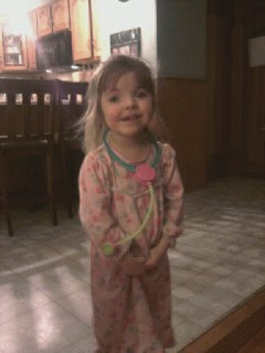
 Dr. Sarah Jeffers is in the house! Sarah has opened up her practice in our playroom, and the waiting room is in the living room. You need to wait your turn until she calls you in. It is quite cute. She pretends to take your blood like she had taken at the hospital this week, and will listen to your heart. FYI.....she is taking new patients!
Tuesday, February 15, 2011
Pre-Op Visit at Children's Hospital
Today I took Sarah for her Pre-Op visit at Children's Hospital. We had to be there for 9am and true to fashion traffic was horrendous but we made it! The first thing Sarah saw was a computer which she happily played with as we waited. It was a busy morning. We met with Nursing, Anesthesia, Admitting, a Plastic Surgery Nurse Practitioner, and finally went to the Laboratory for blood work. Sarah was quite a brave girl as they they took blood. Everything is on schedule for March 1st. We hope and pray Sarah is able to stay healthy over the next 2 weeks so we can proceed as planned. The doctors, nurses, and staff at Children's Hospital continue to be so helpful and kind to Sarah. We feel like Sarah is in good hands. Other than the blood work she had done today, she had a great time coloring and doing activities with the Child Life program at the hospital. As of now Sarah thinks the hospital is a pretty fun place. She left with a balloon and prize in hand.
Wednesday, February 9, 2011
What is Saethre-Chotzen Syndrome
Sarah has what's called Saethre-Chotzen Syndrome. This genetic condition is the cause for the Craniosynostosis. Anyone can be born with Craniosynostosis but those who have this genetic condition are higher at risk. Usually when the craniosynostosis repair is completed subsequent surgeries are not needed; however, because Sarah has Saethre-Chotzen the fusion happens at a higher rate. Saethre-Chotzen syndrome presents itself differently in each person and each family. For example, Jason has the same syndrome but does not have Craniosynostosis. Jason has the ptosis (droopy eyelids), but Sarah doesn't have that. Below is more detailed information about the syndrome:
What is Saethre-Chotzen syndrome?
Saethre-Chotzen syndrome is a genetic condition characterized by the premature fusion of certain skull bones (craniosynostosis). This early fusion prevents the skull from growing normally and affects the shape of the head and face.
Most people with Saethre-Chotzen syndrome have prematurely fused skull bones along the coronal suture, the growth line that goes over the head from ear to ear. Other parts of the skull may be malformed as well. These changes can result in an abnormally shaped head, a high forehead, a low frontal hairline, droopy eyelids (ptosis), widely spaced eyes, and a broad nasal bridge. One side of the face may appear noticeably different from the other (facial asymmetry). Most people with Saethre-Chotzen syndrome also have small, unusually shaped ears.
The signs and symptoms of Saethre-Chotzen syndrome vary widely, even among affected individuals in the same family. This condition can cause mild abnormalities of the hands and feet, such as fusion of the skin between the second and third fingers on each hand and a broad or duplicated great toe. Delayed development and learning difficulties have been reported, although most people with this condition are of normal intelligence. Less common signs and symptoms of Saethre-Chotzen syndrome include short stature, abnormalities of the bones of the spine (the vertebra), hearing loss, and heart defects.
Robinow-Sorauf syndrome is a condition with features similar to those of Saethre-Chotzen syndrome, including craniosynostosis and broad or duplicated great toes. It was once considered a separate disorder, but was found to result from mutations in the same gene and is now thought to be a mild variant of Saethre-Chotzen syndrome.
How common is Saethre-Chotzen syndrome?
Saethre-Chotzen syndrome has an estimated prevalence of 1 in 25,000 to 50,000 people.
What are the genetic changes related to Saethre-Chotzen syndrome?
Mutations in the TWIST1 gene cause Saethre-Chotzen syndrome. The TWIST1 gene provides instructions for making a protein that plays an important role in early development. This protein is a transcription factor, which means that it attaches (binds) to specific regions of DNA and helps control the activity of particular genes. The TWIST1 protein is active in cells that give rise to bones, muscles, and other tissues in the head and face. It is also involved in the development of the limbs.
Mutations in the TWIST1 gene prevent one copy of the gene in each cell from making any functional protein. A shortage of the TWIST1 protein affects the development and maturation of cells in the skull, face, and limbs. These abnormalities underlie the signs and symptoms of Saethre-Chotzen syndrome, including the premature fusion of certain skull bones.
A small number of cases of Saethre-Chotzen syndrome have resulted from a structural chromosomal abnormality, such as a deletion or rearrangement of genetic material, in the region of chromosome 7 that contains the TWIST1 gene. When Saethre-Chotzen syndrome is caused by a chromosomal deletion instead of a mutation within the TWIST1 gene, affected children are much more likely to have intellectual disability, developmental delay, and learning difficulties. These features are typically not seen in classic cases of Saethre-Chotzen syndrome. Researchers believe that a loss of other genes on chromosome 7 may be responsible for these additional features.
Read more about the TWIST1 gene and chromosome 7.
Can Saethre-Chotzen syndrome be inherited?
This condition is inherited in an autosomal dominant pattern, which means one copy of the altered gene in each cell is sufficient to cause the disorder. In some cases, an affected person inherits the mutation from one affected parent. Other cases may result from new mutations in the gene. These cases occur in people with no history of the disorder in their family.
Some people with a TWIST1 mutation do not have any of the obvious features of Saethre-Chotzen syndrome. These people are still at risk of passing on the gene mutation, and may have a child with craniosynostosis and the other typical signs and symptoms of the condition.
What The Surgery Involves and Why?
The team of surgeons working with Sarah are going to perform three procedures the day of Sarah’s surgery. The first is a Craniosynostosis Repair in which they will increase the size of her skull and allow her brain more room to grow. The second is to fill in any gaps in bone. There are lots of little soft spots on Sarah’s skull where bone did not fill in from her previous surgery. They will use pieces of her own bone mixed with another material to fill in these spots and it will generate bone growth. The third procedure is called fronto-orbital advancement. Sarah’s forehead is very flat, pretty much flush with her eyes. They will bring her forehead forward. Below is some information for those that are interested that gives more detail into some of what we have discussed here. If you have any questions please feel free to comment and we will answer them the best we can.
Craniosynostosis
Craniosynostosis is a term that refers to the early closing of one or more of the sutures of an infant's head. The skull is normally composed of bones which are separated by sutures. This diagram shows the different sutures which can be involved.
As an infant's brain grows, open sutures allow the skull to expand and develop a relatively normal head shape. If one or more of the sutures has closed early, it causes the skull to expand in the direction of the open sutures. This can result in an abnormal head shape. In severe cases, this condition can also cause increased pressure on the growing brain.
Types of Craniosynostosis
In sagittal synostosis (scaphocephaly), the sagittal suture is closed. As a result, the infant's head does not expand in width but grows long and narrow to accommodate the growing brain. The sagittal suture is the most common single suture involved in craniosynostosis. The incidence of sagittal synostosis in the population is approximately 1 in 4200 births. Males are affected about three times as often as females.
When the metopic suture is closed, this condition is called metopic synostosis. You may also hear the term trigonocephaly used to describe your child's head shape. The deformity can vary from mild to severe. There is usually a ridge down the forehead that can be seen or felt and the eyebrows may appear "pinched" on either side. The eyes may also appear close together.
The coronal suture goes from ear to ear on the top of the head. Early closure of one side, unilateral coronal synostosis (plagiocephaly) results in the forehead and orbital rim (eyebrow) having a flattened appearance on that side. This gives a "winking" effect. These features may also be more apparent when looking at the child in the mirror.
(This is what Sarah has) Both sides are fused in bicoronal synostosis (brachycephaly). In these cases, the child may have a very flat, recessed forehead. This suture fusion is most often found in Crouzon's, Saethre-Chotzen and Apert's Syndromes.
How is Craniosynostosis diagnosed?
· There are several clues that may have caused you or your doctor to suspect that your child has craniosynostosis. A misshapen head is usually the first clue. The anterior fontanelle, or soft spot, may or may not be open. The suspected diagnosis is confirmed by x-rays. A CT scan is also done to make sure there are no underlying abnormalities in the brain
Craniosynostosis Repair is surgery to fix damage caused by a birth defect that makes the bones in a child’s skull grow together too early.
Alternative Names
Craniectomy; Synostectomy; Strip craniectomy; Endoscopy-assisted craniectomy; Sagittal craniectomy; Frontal-orbital advancement; FOA
Description
A baby's head, or skull, is made up of many different bones. The connections between these bones are called sutures. When a baby is born, it is normal for these sutures to be open a little. This gives the baby’s brain and head room to grow.
Your baby was born with craniosynostosis, a condition that caused 1 or more of your baby’s sutures to close too early. This can cause the shape of your baby’s head to be different than normal. Sometimes it can cause brain damage.
An x-ray or computed tomography (CT scan) can be used to diagnose craniosynostosis. Surgery is usually needed to correct it. This surgery is performed in the operating room under general anesthesia (your child will be asleep and will not feel pain).
Traditional surgery is called open repair. It includes these steps:
The most common place for an incision (a cut made during surgery) to be made is over the top of the head, from just above 1 ear to just above the other ear. The incision is usually wavy. The exact placement of the incision may be different for different problems.
A flap of skin, tissue and muscle below the skin, and the tissue covering the bone are loosened and raised up so the surgeon can see the bone.
A strip of bone is usually removed where 2 sutures connect. This is called a strip craniectomy. Sometimes, larger pieces of bone must also be removed. This is called synostectomy. Parts of these bones may be changed or reshaped while they are outside of the skull and then put back in. Other times, they are removed and not put back in.
Sometimes, bones that are left in place need to be shifted or moved.
Bones are then put into place using a plate with screws that go into the skull.
Surgery usually takes 3 to 7 hours. Your child will probably need to have a blood transfusion during or after surgery to replace blood that is lost during the surgery.
Why The Procedure Is Performed
Surgery frees the sutures that are fused. It also reshapes the brow, eye sockets, and skull as needed. The goals of surgery are:
To relieve any pressure on the child’s brain
To make sure there is enough room in the skull to allow the brain to properly grow
To improve the appearance of the child's head
Risks
Risks for any surgery are:
Breathing problems
Infection, including in the lungs, urinary tract, and chest
Blood loss (children having an open repair may need a transfusion)
Reactions to medicines
Possible risks of having this surgery are:
Infection in the brain
Brain swelling
Damage to brain tissue
After The Procedure
After the open surgery, your child will be taken to an intensive care unit (ICU). After 1 or 2 days, your child will be moved to a regular hospital room. Your child will stay in the hospital for 3 to 7 days.
Your child will have a large bandage wrapped around their head. They will also have an IV (a tube that goes into their vein). The nurses will watch your child closely.
Tests will be done to see if your child lost too much blood during surgery. The doctor may give your child blood through a transfusion if they need it.
Your child will have swelling and bruising around their eyes and face. Sometimes, their eyes may be swollen shut. This often gets worse in the first 3 days after surgery, but it will be better by day 7.
Your child should stay in bed for the first few days. The nurses will keep the head of your child’s bed raised to help keep down swelling.
Talking and singing to the child, and playing music and telling stories, may help soothe them. Acetaminophen (Tylenol) is used for pain, but your nurse will have other pain medicines if your child needs them.
Most children who have endoscopic surgery can go home after staying in the hospital 1 night.
Outlook (Prognosis)
Most times, craniosynostosis repair is successful and allows your child’s skull and brain to develop normally.
Monday, February 7, 2011
Subscribe to:
Comments (Atom)