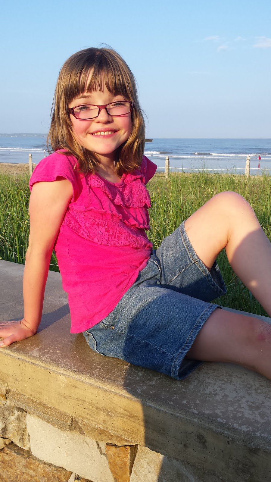
The team of surgeons working with Sarah are going to perform three procedures the day of Sarah’s surgery. The first is a Craniosynostosis Repair in which they will increase the size of her skull and allow her brain more room to grow. The second is to fill in any gaps in bone. There are lots of little soft spots on Sarah’s skull where bone did not fill in from her previous surgery. They will use pieces of her own bone mixed with another material to fill in these spots and it will generate bone growth. The third procedure is called fronto-orbital advancement. Sarah’s forehead is very flat, pretty much flush with her eyes. They will bring her forehead forward. Below is some information for those that are interested that gives more detail into some of what we have discussed here. If you have any questions please feel free to comment and we will answer them the best we can.
Craniosynostosis
Craniosynostosis is a term that refers to the early closing of one or more of the sutures of an infant's head. The skull is normally composed of bones which are separated by sutures. This diagram shows the different sutures which can be involved.
As an infant's brain grows, open sutures allow the skull to expand and develop a relatively normal head shape. If one or more of the sutures has closed early, it causes the skull to expand in the direction of the open sutures. This can result in an abnormal head shape. In severe cases, this condition can also cause increased pressure on the growing brain.
Types of Craniosynostosis
In sagittal synostosis (scaphocephaly), the sagittal suture is closed. As a result, the infant's head does not expand in width but grows long and narrow to accommodate the growing brain. The sagittal suture is the most common single suture involved in craniosynostosis. The incidence of sagittal synostosis in the population is approximately 1 in 4200 births. Males are affected about three times as often as females.
When the metopic suture is closed, this condition is called metopic synostosis. You may also hear the term trigonocephaly used to describe your child's head shape. The deformity can vary from mild to severe. There is usually a ridge down the forehead that can be seen or felt and the eyebrows may appear "pinched" on either side. The eyes may also appear close together.
The coronal suture goes from ear to ear on the top of the head. Early closure of one side, unilateral coronal synostosis (plagiocephaly) results in the forehead and orbital rim (eyebrow) having a flattened appearance on that side. This gives a "winking" effect. These features may also be more apparent when looking at the child in the mirror.
(This is what Sarah has) Both sides are fused in bicoronal synostosis (brachycephaly). In these cases, the child may have a very flat, recessed forehead. This suture fusion is most often found in Crouzon's, Saethre-Chotzen and Apert's Syndromes.
How is Craniosynostosis diagnosed?
· There are several clues that may have caused you or your doctor to suspect that your child has craniosynostosis. A misshapen head is usually the first clue. The anterior fontanelle, or soft spot, may or may not be open. The suspected diagnosis is confirmed by x-rays. A CT scan is also done to make sure there are no underlying abnormalities in the brain
Craniosynostosis Repair is surgery to fix damage caused by a birth defect that makes the bones in a child’s skull grow together too early.
Alternative Names
Craniectomy; Synostectomy; Strip craniectomy; Endoscopy-assisted craniectomy; Sagittal craniectomy; Frontal-orbital advancement; FOA
Description
A baby's head, or skull, is made up of many different bones. The connections between these bones are called sutures. When a baby is born, it is normal for these sutures to be open a little. This gives the baby’s brain and head room to grow.
Your baby was born with craniosynostosis, a condition that caused 1 or more of your baby’s sutures to close too early. This can cause the shape of your baby’s head to be different than normal. Sometimes it can cause brain damage.
An x-ray or computed tomography (CT scan) can be used to diagnose craniosynostosis. Surgery is usually needed to correct it. This surgery is performed in the operating room under general anesthesia (your child will be asleep and will not feel pain).
Traditional surgery is called open repair. It includes these steps:
The most common place for an incision (a cut made during surgery) to be made is over the top of the head, from just above 1 ear to just above the other ear. The incision is usually wavy. The exact placement of the incision may be different for different problems.
A flap of skin, tissue and muscle below the skin, and the tissue covering the bone are loosened and raised up so the surgeon can see the bone.
A strip of bone is usually removed where 2 sutures connect. This is called a strip craniectomy. Sometimes, larger pieces of bone must also be removed. This is called synostectomy. Parts of these bones may be changed or reshaped while they are outside of the skull and then put back in. Other times, they are removed and not put back in.
Sometimes, bones that are left in place need to be shifted or moved.
Bones are then put into place using a plate with screws that go into the skull.
Surgery usually takes 3 to 7 hours. Your child will probably need to have a blood transfusion during or after surgery to replace blood that is lost during the surgery.
Why The Procedure Is Performed
Surgery frees the sutures that are fused. It also reshapes the brow, eye sockets, and skull as needed. The goals of surgery are:
To relieve any pressure on the child’s brain
To make sure there is enough room in the skull to allow the brain to properly grow
To improve the appearance of the child's head
Risks
Risks for any surgery are:
Breathing problems
Infection, including in the lungs, urinary tract, and chest
Blood loss (children having an open repair may need a transfusion)
Reactions to medicines
Possible risks of having this surgery are:
Infection in the brain
Brain swelling
Damage to brain tissue
After The Procedure
After the open surgery, your child will be taken to an intensive care unit (ICU). After 1 or 2 days, your child will be moved to a regular hospital room. Your child will stay in the hospital for 3 to 7 days.
Your child will have a large bandage wrapped around their head. They will also have an IV (a tube that goes into their vein). The nurses will watch your child closely.
Tests will be done to see if your child lost too much blood during surgery. The doctor may give your child blood through a transfusion if they need it.
Your child will have swelling and bruising around their eyes and face. Sometimes, their eyes may be swollen shut. This often gets worse in the first 3 days after surgery, but it will be better by day 7.
Your child should stay in bed for the first few days. The nurses will keep the head of your child’s bed raised to help keep down swelling.
Talking and singing to the child, and playing music and telling stories, may help soothe them. Acetaminophen (Tylenol) is used for pain, but your nurse will have other pain medicines if your child needs them.
Most children who have endoscopic surgery can go home after staying in the hospital 1 night.
Outlook (Prognosis)
Most times, craniosynostosis repair is successful and allows your child’s skull and brain to develop normally.

1 comment:
Sarah- we are thinking about you and saying some prayers! You are adorable. and all good wishes to Bill, Jill and Jason during this very tough time.
Janet, Don , Emily and Jessica Cann
Post a Comment